Overview
Conservators use several kinds of examination and imaging techniques, some which require specialized equipment capable of capturing light wavelengths that are invisible to the naked eye, to understand the many layers of an artwork.
These imaging techniques can give conservators more information about the techniques the artist used to create the object, the artist's material choices, and the history of the object. This information helps determine the best treatment option for objects, and also to provide institutions and collectors with art historical information about their artworks. Below are a few of the state-of-the-art techniques BACC conservators use to analyze artworks.
These imaging techniques can give conservators more information about the techniques the artist used to create the object, the artist's material choices, and the history of the object. This information helps determine the best treatment option for objects, and also to provide institutions and collectors with art historical information about their artworks. Below are a few of the state-of-the-art techniques BACC conservators use to analyze artworks.
Ultraviolet Light
One of the first non-destructive imaging tools conservators use to examine an artwork is ultraviolet light (UV). Ultraviolet light is just beyond violet in the visible spectrum (just below 400 nm); it's the same light we protect our skin from with sunblock. It is generally used to look at the surface of a painting. With UV light, we can often see aged varnishes because they fluoresce or glow. The color of the fluorescence can help indicate which kind of varnish or coating was used on the surface. UV light can also show areas of retouching or old, painted restoration.
Infrared Reflectography
Infrared reflectography, which uses energy just beyond the visible light spectrum in the opposite direction (above red at 700 nm), is often employed for the detection of preparatory underdrawing lines against a light-colored background. Infrared light can penetrate or see through some top paint layers and reveal other paint layers or drawings below. Infrared reflectography, which is also a non-destructive imaging technique, has mainly been used in the past for the examination of elaborate underdrawings in Renaissance-era paintings from northern Europe and Italy.
|
|
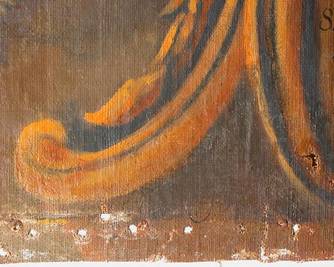
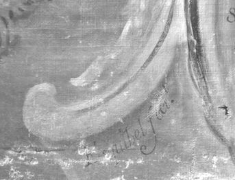
Infrared reflectography can also be used to see through later additions of paint, such as in this late 18th century Spanish Colonial painting shown above. The IR reflectogram revealed “Esquibel fecit” (see above, right). Jose Joachim Esquibel was known to have a workshop near Mexico City in the late 18th century. With infrared reflectography, conservators were able to discover the artist of this Last Judgement scene.
X-Radiography
X-radiography, the same technique used to look at broken bones in the human body, is also used to see inside of paintings. X-radiography uses energy found beyond ultraviolet light in the electromagnetic spectrum and can penetrate deeper into the layers of an object. X-radiographic detectors record the differences in densities in a painting; dense areas appear white on an X-ray because the denser materials contained in an object impede the penetration of X-rays. Essentially, X-radiography is a record of the varying densities in the painting. Thick wood appears whiter than surrounding areas because it is physically denser, while lead white paint appears whiter because it is chemically denser than other paints. With X-rays, conservators can see paint losses, tears, or changes made to the painted composition. This non-invasive technique can also help with looking at canvas weave and the construction of wooden panels. All of this examination helps us to understand the painting's construction, material history, and its condition.

|
|
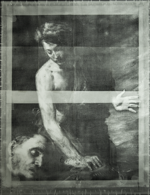
Above, left, we can see the X-ray of a panel painting supported by an elaborate wooden auxiliary system called a cradle (the wooden grid). The white spots are remnants of the original, more simplified, wooden auxiliary support system; this information helps conservators understand how the panels were originally held together. Above, right, the X-ray of the portrait on canvas shows the canvas's weave, a tear in the canvas at the bottom of the painting, the tacks used to secure the canvas to the stretcher, and the lead white passages of paint used in the face.
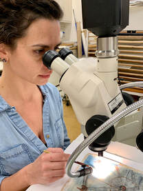
Portable X-Ray Fluorescence
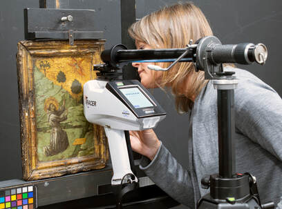 Using the X-Ray Fluorescence Spectrometry (XRF) device.
X-Ray Fluorescence Spectrometry (XRF) is a non-destructive technique for elemental analysis. The XRF device is placed in front of an unknown substance, and the sample is bombarded with high energy x-rays causing the atoms to emit characteristic secondary or fluorescent x-rays that are reflected back into the XRF detector. The resulting spectra can be analyzed to determine which elements are present in the unknown. Knowing the elemental composition of an unknown, such as pigment within a paint film, can help determine which pigments are present. For example, if a red area shows the presence of mercury (Hg), this would be consistent with the presence of the pigment vermilion (mercuric sulfide.) This technique can measure the presence of elements heavier than sodium and is most useful for inorganic pigments and metals.
POLARIZED LIGHT MICROSCOPY
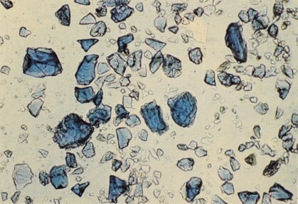 Dispersed azurite pigment particles as viewed under magnification.
Polarized-light microscopy is used to identify the individual pigment particles that make up paint layers. This technique requires that extremely small samples be taken from the surface of the painting.
A small scraping of the top layer of paint is mounted on a microscope slide and viewed under a microscope with light polarizing capabilities. The pigment particles are examined under high magnification to determine standard particle characteristics, such as color, morphology, transparency, birefringence/isotropism, index of refraction, particle relief, pleochroism, polarization colors, and particle size.
It is possible to identify the pigments in the sample by knowing the particular characteristics and then comparing them with the characteristics of reference pigment samples. This technique should be confirmed with elemental analysis, whenever possible, to make a conclusive identification.
Polarized-light microscopy can also be used in the characterization and identification of fibers used to make canvas and paper. This technique requires that extremely small samples be taken. The sample is mounted on a microscope slide and viewed under a microscope with light polarizing capabilities. The fibers are examined under high magnification to determine the morphology of the fibers, and they can be compared to reference slides of known fibers.
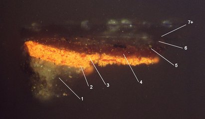
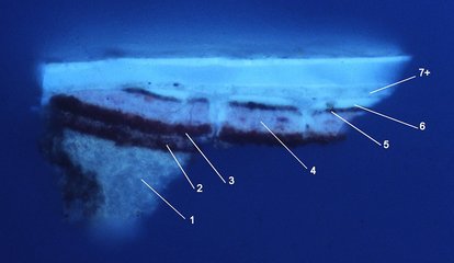
Cross-Sections
Cross-section microscopy is the examination of the layering structure of paintings. Cross sections can yield information about artistic technique, condition, and material content that cannot be ascertained by viewing the surface of a painting alone. This technique requires that a microscopic sample be removed from the painting, embedded in resin, polished along its depth, and then examined under a microscope. The sample should include the full stratigraphy of a painting, from topmost surface to ground layer or substrate. Micro-chemical staining can be performed on a cross-section to characterize and locate oil, gum, or protein in binding media which can be instructive for contextualizing a painting stylistically and/or historically.
|
|
Above, left shows a cross section of a shadow of a red robe from a historic panel painting in ordinary, visible light. Above, right shows the same cross section in ultraviolet light. Original magnification 400x. Key: 1) gesso ground, (2) and (3) two layers of vermilion, (4) red glaze, (5) thin light brown layer, (6) and (7) the varnish layers can be seen in ultraviolet light. Layer (6) can be seen extending into the cracks in the paint, indicating that it is not an original varnish coating.